

POSTER ABSTRACTS
Materials should NOT be shared with those that are not registered for the conference. Poster abstracts are not proofed for spelling and/or grammar errors.
The poster and/or other information contained on this website may NOT be downloaded and/or used without prior written permission from all authors on the project. If you would like to be connected with the author(s), please email [email protected].
Chronic intermittent hypoxia adversely affects renal microcirculatory regulation and tissue PO2 in ovariectomized female rats
Raina M. Gerritts, DO’241, Benjamin G. Madigan, DO’241, Katherine A. Harbeck, DO’241, Kelsey S. Schwartz2, James A. Lang, PhD2, Abbie A. Voas, DO’24, MSA’221, Sarah C. Clayton, PhD1, and Noah J. Marcus, PhD1
1College of Osteopathic Medicine, Des Moines University, Des Moines, IA
2Department of Kinesiology, Iowa State University, Ames, IA
Abstract
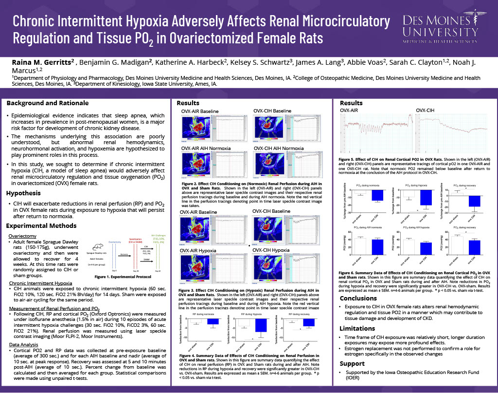
Sleep apnea is highly prevalent in post-menopausal women and is a major risk factor for chronic kidney disease. Abnormal renal hemodynamics and hypoxemia likely play prominent roles in this process. We sought to determine if chronic intermittent hypoxia (CIH, a model of sleep apnea) would adversely affect renal perfusion (RP) and PO2 in ovariectomized (OVX) rats. We hypothesized that CIH would exacerbate reductions in RP and PO2 in OVX rats during exposure to hypoxia that would persist after return to normoxia. Adult female OVX Sprague Dawley rats (n=4-5 per group) were randomized to 14 days of CIH or sham. Regulation of RP and PO2 was assessed under isoflurane anesthesia (1.5% in air) during 10 episodes of hypoxia (AIH, FiO2 10%, FiCO2 3%). RP was measured using laser speckle contrast imaging and PO2 was measured using fiber optic probes (Oxford Optronix). Renal cortical tissue was assessed for expression of eNOS mRNA (qRT-PCR) and protein (western blot). RP and cortical PO2 were significantly decreased in both groups during both normoxia and hypoxia portions of AIH relative to baseline (p<0.05), but to a greater extent in OVX-CIH animals (p<0.05). Reductions in cortical PO2 during normoxia remained below baseline at 5 minutes post-AIH (p<0.05). Expression of eNOS was decreased in OVX animals relative to intact females but was not different between OVX-AIR and OVX-CIH. In conclusion, exposure to CIH in OVX female rats alters renal hemodynamic regulation and tissue PO2 in a manner which may contribute to tissue damage and development of CKD.
Access PDF version to expand view.